This was a 16 month male patient with intermitent constipation with a palpable pelvic mass that initially was thought to be a dilated sigmoid but later pediatrician noticed that the mass had smooth edges and had a circular configuration. An ultrasound was made (not shown) that reported distended bowel loops hence difficulted the correct visualization, although a pelvic heterogeneous mass was seen mostly echoic with acoustic shadowing but barely depicted. Padiatrician recommended a CT scan, here are the images:


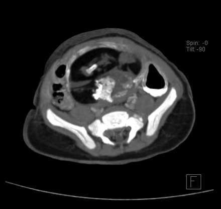


Findings and differentials??...later on I'll post the conclusion of this case.
Este comentario ha sido eliminado por el autor.
ResponderEliminarLarge dermoid!
ResponderEliminar