a.

In here (Image a.)we visualize some amount of free fluid in the posterior renal fossa wich is very important to recognize in a post-trauma patient because it could be hematoperitoneum and we as radiologists need to find the cause of it such as visceral or vascular trauma.
b.

Then (Image b.) this round hiperecogenic image is seen in the right adrenal gland that has the appearance and ecogenicity of an adenoma so therefore, we suggested a CT scan.
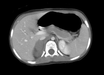
c.

In these images we visualize the lesion and with IV contrast we see that has barely enhancement...almost none enhancement one might say...if you look closer you can see that first of all, it has no fat density in order to think of an adenoma and that there is some stranding of IV contrast into abdomen.But here comes the tricky part, what if this was an incidental finding and in fact, we are looking an adenoma that was there long before the trauma happened??...is this finding clinically significant??
First of all, we should rely on history background and some clinical data. This is a pediatric trauma patient who has moderate abdominal pain with suspected visceral trauma so hematoma of any organ should be on top of my differentials and then exclude another posibilities. Rest of physical exam is normal. With this in mind and preliminary CT and US findings, 2 questions crossed my mind:
- Is adrenal hematoma is an urgency?..
Literature says that adrenal hematoma is very uncommon and therefore is poorly characterized. A 1992 review published in AJR says that eventhough is more common in pediatric population than in adults, adrenal hemorraghe has a 3% of incidence, is unilateral in 85% of cases and with strong predominance of right gland. In other article published in Journal of Urology states that adrenal hematoma ussually comes as a part in a multyorgan trauma but if unilateral, can be self-limited and requires no surgical or intensive care unit treatment. Dr. Fishman on his web resource CTisus describes adrenal imaging as well as epidemiology and states that eventhough is important to diagnose adrenal hematoma, the real deal is when bilateral and there it becomes a lifethreatning condition with high mortality rates. This takes me to my second question:
- How is the radiological presentation of adrenal hematoma?..
Now, this is a challenging question to answer because of the very low incidence and variable spectrum. Dr. Fishman also makes a beautiful description on how is the imaging presentation of adrenal hematoma especially on CT. First, the most common presentation we see is an enlarged adrenal gland with a cyst in appearence but has IV enhancement. He adverts, often we see also barely enhanced adrenal hematomas ussually in the acute setting. Here are some examples of poor enhancement:

March 2004 Radiology, 230,669-675.

If our next differential is adenoma, how are the imaging characteristics?
Again, what we find very helpful in this case is the epidemiology factor. How often we see adenomas in infants?. What other clinical characteristics we need to look up to to suspect that in fact is an adenoma?. The imaging characteristics as we know are almost the same as an adrenal non enhanced hematoma. The main resource we have is to characterize the lesion according to the % of washout of IV contrast as proposed by Dr Caoili. To my concern I don't know if this is used also in pediatric patients. But before we propose this, we have to keep in mind that adrenal hyperplasia is more common than adenomas in children and when suspected has malignant potential and are functional wich means that develops hormonal disturbance with clinical manifestations. So in this case, adenoma is less likely.
Patient had conservatory approach and with 2 days of monitoring, he was sent home with no complications.
Again, fell free to comment...thank you!
Sivit, C.J., Ingram, D.J., Taylor, G.A., Bulas, D.I., Kushner, D.C., Eichelberger, M.R. - Posttraumatic adrenal hemorrhage in children: CT findings in 34 patients. AJR Am. J. Roentgenol, 1992, 158:1299.
Gabak-Shehab L, Alagiri M. Traumatic adrenal injuries J Urol. 2005 Apr;173(4):1330-1.
Caoili EM, Korobkin M, Francis IR, Cohan RH, Platt JF, Dunnick NR, Raghupathi KI.Adrenal masses: characterization with combined unenhanced and delayed enhanced CT.Radiology. 2002 Mar;222(3):629-33.
No hay comentarios:
Publicar un comentario