jueves, 18 de julio de 2013

CASE 37: 16 m/o male patient with a palpable mass

Here is a case in which Radiology plays an important role in management. This case was provided by Tecniscan Diagnostic Center.

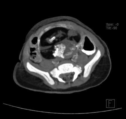
This was a 16 month male patient with intermitent constipation with a palpable pelvic mass that initially was thought to be a dilated sigmoid but later pediatrician noticed that the mass had smooth edges and had a circular configuration. An ultrasound was made (not shown) that reported distended bowel loops hence difficulted the correct visualization, although a pelvic heterogeneous mass was seen mostly echoic with acoustic shadowing but barely depicted. Padiatrician recommended a CT scan, here are the images:


Findings and differentials??...later on I'll post the conclusion of this case.

lunes, 1 de julio de 2013

DX: Cystic brain metastases from lung cancer

Cystic lesions in brain have a broad spectrum. In order of frecuency, NCC is a parasitic infection common in  this lattitude so, we recommended pharmacological treatment and a new MR control when over. Patient did not resolve symptoms and control (not shown) was without change.  In efforts to a definitive diagnosis, we called the neurologist to review patient history. We found that this patient was treated years ago from lung small cell carcinoma!. He underwent chemotherapy and was in remission. He was complaining from cough and chest pain since neurological symptoms. Surprisingly, he skipped the yearly CT control in the past year. We asked for a thorax CT control. 

Here is the control wich clearly shows a left perihiliar lung mass with inflitration to thoracic wall:


With this in mind the possibility of a metastases was clear, even though cystic metastases are uncommon. Brain glioma and abscess were good differentials as well although DWI was completely normal, so abscess can be almost ruled out. We recommended a Magnetic Resonance Spectroscopy (in other institution), shown above:


Single-Voxel MRS showed increased lactate peak and reduction in NAA/Cr and Cho/Cr. Elevation in lactate are found in high-grade gliomas and metastases. Due to medical background metastases was  considered. Almost all high grade gliomas are found with elevation in NAA or Cho metabolites. In summary, although MRS can give information of the celularity of the tumor and in consecuence narrow the differentials, clinical data are mandatory in confirming the diagnosis. Patient is programmed to have a biopsy but oncologic therapy was initiated.

Detection of an intracranial mass on imaging in patients with a known primary raises the possibility of metastasis. Often the diagnosis of an intracranial metastasis is made radiologically without histological confirmation. However, radiological evaluation has its own limitation. Though the MRI has increased the specificity of diagnosis, routine MRI sequences may still not be able to differentiate between different intracranial lesions. The classical appearance of a metastasis is a solid enhancing mass with well-defined margins and extensive edema. Occasionally, central necrosis produces a ring enhancing mass. Such a lesion is difficult to differentiate from an abscess. Recently, diffusion and spectroscopy sequences have proven useful to differentiate between the two entities.

Brain metastases occur in 25%-35% of all cancer patients. The majority of brain metastases are solid tumors; cystic brain metastases are unusual. Several reports of cystic brain metastases associated with non-small cell lung carcinoma have been described, but small cell lung carcinoma presenting with cystic brain metastases is previously recognized in only one study.


This was a difficult case and I appreciate your comments. As some might have seen I'm no expert in MRS yet  :) . All comments are welcome. 


Here are some useful links, 


miércoles, 26 de junio de 2013

CASE 36: Mid 50's patient with aphasia

This case had a great interdisciplinary approach result from a good communication with the Neurology Department and other Departments also.

Patient reffered problems with understanding the meaning of words associated with progressive headache that started 2 months ago. He did not suffered from any other neurological symptoms. He was directly reffered to us to undergo an MR with suspicion of a mass or a stroke onset.

Here are the relevant images:


FLAIR


 T1


POST GAD


T2


With this medical history given, make some differential diagnosis. Later on, I'll post further information and conclusion.
  

jueves, 20 de junio de 2013

DX: Hepatic Encephalopathy (Hyperammonemia)

Patient had chronic Hepatitis B infection with chirrosis. Ammonia levels were high and was having an hepatic failure. Patient symptoms were mild several months ago but were progressively worsening.

She was classified in Stage 3 of the West Haven criteria for Hepatic Encephalopathy

MRI findings are unspecific and we ussually see this kind of cortical T2 hyperintensities in various forms of encephalitis. It had no restriction in DWI and T1 was completely normal. Clinical history is imperative to make this diagnosis.

In this case, the characteristic imaging feature is the symmetric thalamic altered signal mainly in the pulvinar regions. Usually, hepatic encephalopathy affects globus pallidus such as this case also. Other clue is the cortical and subcortical hyperintensities that may reflect edema because of swelling of the astrocytes. These are often described that mimic ALS (Lou Gehrig's).

Here are the relevant images: (sorry the poor quality but we work with 1T Magnet).


Here is a similar case on literature:

AJNR 2010 31: 1471-1479


There is a debate on wheter ammonia levels correlate with the onset of Encephalopathy. There are plenty litereture on this subject that goes beyond the scope of this blog but it's interesting to check it out.

Here are some useful links:

http://www.ajnr.org/content/29/9/1612.full.pdf+html
http://www.ajnr.org/content/31/8/1471.full.pdf+html
http://emedicine.medscape.com/article/186101-overview
http://www.aafp.org/afp/2003/1001/p1408.html


Thank you Dr. Muñoz, Dr. Gurrala Ravikiran and Dr. Prabhu Radhan for your participation in the discussion of this case.

Until next time!


domingo, 16 de junio de 2013

CASE 35: Altered mental status

Hello again!..long time.

This case is a female patient with sudden altered mental status. She was previously admitted to the Hospital with history of fever, nausea, vomiting and headaches. She had congestive heart failure and HBV infection.

Here are the images:


Findings?..DD?..Soon the conclusion.


domingo, 12 de mayo de 2013

GONE FOR A FEW WEEKS

Gonna be out for a few couple of weeks perhaps a month or so...I'm currently doing my MSK rotation and later I'll conclude a Course of Medical Neuroscience. I know it's not Radiology field completely but it's very helpful and I'm happy learning it...I'll post you soon!..thanks.


Sincerely,

Edgar J. Salguero, MD
PGY4-Chief Resident

lunes, 22 de abril de 2013

DX: Olfactory groove meningioma

This case shows some of the peculiar imaging characteristics of a Meningioma. It was hyperdense on CT post contrast, T1 and T2 iso-hyperintense, and hyperintense on FLAIR images with avid enhancement on post Gad. It had a dural based tail and showed a falx calcification. It was located in the orbitofrontal region just above the cribiform plate and near the midline. It had vasogenic edema.

Here are the images:


Although the tumor is small in size it provoked considerable presence of edema that probably caused symptoms. These symptoms were vague with components of a frontal lobe disorder that could explain changes in personality. There was no Anosmia that could lead us to the specific location of symptoms. But current literature says that symptoms may vary in this condition. This tumor is benign but has great growing potential so diagnosis is crucial.

Olfactory groove meningiomas arise over the cribriform plate and can be differentiated from tuberculum sellae meningiomas because olfactory groove meningiomas arise more anterior in the skull base and displace the optic nerve and chiasm inferiorly rather than superiorly. Although these tumors arise in the midline, they may extend predominantly to one side like this case. It can also be very atypical in presentation with restricted DWI.


Thank you for your FB comments, you can participate here as well.


http://www.medscape.com/viewarticle/457741
http://radiopaedia.org/cases/olfactory-groove-meningioma
http://www.neuroradiologycases.com/2011/10/olfactory-groove-meningioma.html