domingo, 8 de julio de 2012

DX: Left Carotid Artery Loop or Coil. Vertebral artery artifact

Though this case is incomplete and had technical deficiencies (such as: 1 Tesla Magnet; TOF sequence; no arteriogram; and lack of showing aortic arch and carotid and subclavian proximal segments). All of this issues makes diagnosis a little difficult.

First of all, TOF sequence itself always has its ups and downs because of low sensitivity. In addition, 3D reconstruction predisposes more artifacts. Many of these artifacts can cause pseudostenosis or segmental blurring. This has to be expected when looking at this sequence. To be assured, lets see the raw data previous 3D reformat.


There is an irregular trajectory of the left carotid artery in the upper cervical portion previous to petrous segment. This makes the diagnosis of a Carotid "Loop" more plausible. Clinical symptoms support this posibility. On the other hand, we did not see an abnormal left vertebral artery, that was shown in the 3D virtual reconstruction.


In this case, and arteriogram had to be done so it was reffered to another institution. (Finantial issues).

Carotid Loop or Coil:

The association between kinking or coiling of the internal carotid artery (ICA) and cerebrovascular insufficiency was first noted in 1951. Since then several reports have dealt with the clinical relationship between carotid elongation and kinking and cerebrovascular disease. Although conclusive evidence linking the two is still lacking, certain inferences can now be drawn.

Looping and kinking of the ICA has been observed in infants and even in fetuses. The cause of these loops is related to embryological development. The vessel is formed from the third aortic arch and from the dorsal aorta; hence, in the embryo it is normally kinked.
Straightening occurs when the fetal heart and large vessels recede in the thoracic cavity. If the embryological state persists, it produces different kinds of undulations, loops, and kinks. This anomaly generally does not become symptomatic until later in life.

There appears to be no relationship between the severity of the kinking and either rising blood pressure or increasing age. The role of degenerative changes in the vessel wall remains uncertain. If this factor were important, then kinks should be more severe in older individuals because degenerative changes increase with age; however, this is not generally the case. At best the cause for looping and kinking of the carotid artery may be ascribed partly to embryological development and partly as secondary to atherosclerotic changes.

http://www.crcnetbase.com/doi/abs/10.1201/9780203912904.ch54
http://stroke.ahajournals.org/content/6/6/649.full.pdf
http://bubbasoft.org/carotid_collaterals/carotid_loops.htm


All coments are welcome...have a nice day.

sábado, 30 de junio de 2012

CASE 22: Pt with chronic recurrent brain stroke

This is an easy one I guess but often underestimated as a differential. Patient underwent Neck Angioresonance with a TOF sequence to discard stenosis. Previous carotid Doppler revealed a bilateral non-significant stenosis due to chronic plaque in carotid bulbs.

Here are the 3D VR images; later on I'll post some raw axial slices to confirm findings.

Click on the first image to enlarge:


viernes, 29 de junio de 2012

DX: Acute pontine-mesencephalic infarct

Patient needed to be examined thoroughly by a neurologist because a lesion in this region could have multiple clinical manifestations. First of all, MR gave us a big hint on the nature of the disease: acute infarct. Now where exactly is the lesion? is it pontine or mesencephalic?.. the importance of this is because the origin of some cranial nerves.

Here is the depictive MR:


Maybe it is a borderline lesion between pontine and mesencephalic altough with more extension to cephalic portion of pons.

Good neurological examination could have given us a hint.

Cranio-facial autonomic symptoms such as ipsilateral lachrymation, rhinorrhoea, conjuctival congestion, ptosis, or eyelid oedema are the sine qua non of trigeminal autonomic cephalalgia. It is thought that activation of the trigeminal afferent system generates pain, and co-activation of the VIIth nerve efferent parasympathetic pathway produces the autonomic manifestations such as ipsilateral lachrymation and rhinorrhoea.

Ischaemia in the ponto-mesencephalic junction could involve the trigeminal main sensory and mesencephalic nuclei, resulting in facial dysaesthesia comparable to ischaemic nerve pain. In contrast to the “boring” intense pain often encountered with trigeminal autonomic cephalalgias, the burning dysaesthesias are more consistent with ischaemia. Ischaemia could also trigger trigemino-facial synapses in the superior salivatory nucleus and switch on the facial efferent parasympathetic pathway, resulting in ipsilateral lachrymation and rhinorrhoea.

In this case hemiparesia and hemiplegia were the main symptoms so Millard-Gubler and Raymond-Foville are to consider although the location is usually in medulla or base of the pons.

Named Brainstem Syndromes

EponymSiteCranial NervesTractsSignsUsual Cause
WeberBase of MidbrainIIICorticospinalOculomotor palsy with crossed hemiplegiaVascular, tumor
ClaudeMidbrain tegmentumIIIRed nucleus and Brachium ConjunctivumOculomotor palsy with contralateral cerebellar ataxia and tremorVascular, tumor
BenedictMidbrain tegmentumIIIRed nucleus, corticospinal tract, brachium conjunctivumOculomotor palsy, contralateral cerebellar ataxia, corticospinal signsVascular, tuberculoma, tumor
NothnagelMidbrain tectumUnilateral or bilateral IIISuperior cerebellar pedunclesOcular palsies, paralysis of gaze, cerebellar ataxiaTumor
ParinaudDorsal Midbrain  Paralysis of upward gaze and accommodation, fixed pupils, retraction nystagmusPinealoma, hydrocephalus
Millard-Gubler and Raymond-FovilleBase of PonsVII and sometimes VICorticospinal tractFacial and 6th palsy, contralateral hemiplegia, sometimes gaze palsyVascular,tumor
AvellisMedulla tegmentumXSpinothalamic, sometimes pupillary fibersParalysis of soft palate and vocal cord and contralateral hemianesthesiaInfarct or Tumor
JacksonMedulla TegmentumX,XIICorticospinalAvellis plus ipsilateral tongueInfarct or Tumor
WallenbergMedulla, lateral tegmentumSpinal V,IV,X,XILateral STT,Descending Pupil fibers, Spinocerebellar and olivocerebellar tractsIpsi V, IV, X, XI palsy, Horner's, cerebellar ataxia. Contra pain and tempVascular - Pica or vertebral

 http://www.dizziness-and-balance.com/disorders/central/strokes/brainstem%20strokes.htm


 Check out this articles also:

http://jnnp.bmj.com/content/76/7/1041.1.full
http://en.wikipedia.org/wiki/Lateral_pontine_syndrome
http://www.strokecenter.org/professionals/stroke-diagnosis/stroke-syndromes/inferior-medial-pontine-syndrome-foville-syndrome/
http://www.strokecenter.org/professionals/stroke-diagnosis/stroke-syndromes/ventral-pontine-syndrome-millard-gubler-syndrome/
http://stroke.ahajournals.org/content/28/3/649.full


Hope it was helpful, until next time!

viernes, 22 de junio de 2012

CASE 21: 62 y/o male with altered mental status and focalization

Patient was having hemiparesia and at the time of admission. Here is the CT scan with the relevant finding to my concern:


I suggested an MRI..


Findings and DD please..

miércoles, 13 de junio de 2012

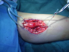
DX: Schwannoma of the upper limb (preliminary)

This is the conslusion of the case. Patient underwent MR because of  US findings consistent with a heterogeneous hypoecoic mass with partial vascularity, sharp edges, within triceps  and biceps brachii muscles along the median or ulnar nerve in the medial plane of the upper arm.  Long axis shows that it presumptively comes from neural origin because fibers attached in cephalic and caudal position of the mass.


MR shows displacement of brachial artery and vein so neural origin has to come in consideration. It is moderate iso-hyperintense on T1 with moderate Gd-DTPA uptake.


Here are some pictures I' ve found on a quick websearch (I do not own this pictures):


http://www.ultrasoundcases.info/Slide-View.aspx?cat=438&case=1966


http://www.jortho.org/2007/4/2/e37/index.htm

 Schwannoma, also known as neurilemmoma, is a benign soft tissue tumour arising from the schwann cells of the nerve sheath. Schwannomas are usually solitary and encapsulated.

Is one of the few truly encapsulated neoplasms of the human body and is almost always solitary. It’s most common locations are the flexor surfaces of the extremities, neck, mediastinum, posterior spinal roots, and cerebellopontine angle. The nerve of origin often can be demonstrated in the periphery, flattened along the capsule but not penetrating the substance of the tumor. Since this is a benign neoplasm every attempt should be made to preserve the nerve.

The Schwannoma (neurilemmoma) is the most common tumor of the peripheral nerve, it accounts for 8% of all primary intracranial tumors and 80-90% of those in the cerebellopontine angle. The peak incidence is in the third to sixth decades, with a slight female predominance. Intracranially there is a predilection for sensory nerves especially the vestibular branch of the eighth nerve. Rarely, schwannomas occur intraparenchymally within the brain, cerebellum, or spinal cord: in such rare instances, they presumably arise from schwann cells that accompany blood vessels.

..

This was an outpatient and was reffered to a Peds Surgeon in other hospital.

viernes, 8 de junio de 2012

CASE 20: 12 y/o girl with left arm palpable mass

This case remarks the importance of the ultrasound. Patient came with history of palpable mass in the medial aspect of distal arm. Physician suspected an abscess but no recent trauma or infection whatsoever.

Sorry for the poor quality but in this US device the only way I could extract images was through a discontinued diskette (yep, old model). Anyways here are the US images:


Ignore the anatomic reference (right rather than left side)


I suggested an MR due to US findings, so here are the relevant slices:


Findings and differentials of both studyes please...soon, the conclusion.

martes, 29 de mayo de 2012

DX: Esophageal calcification after corrosive injury

Sorry for the delay. This couple of months have been quite diffucult but we are still making some noise. Anyways here is the conclusion of this case.

We inquire if patient had a history of caustic ingestion (sodium hydroxide) and he did a couple of months ago. He accidentally swallowed this corrosive but induced himself vomiting shortly after ingestion.

Relevant findings are as described:


There was no adenopathy or infiltration to surrounding tissues.

Primary DD is Leiomyoma.

The course of corrosive damage to the intestinal tract can be divided in three phases:

1) 1.4 days after ingestion, acute necrosis with intensive inflamatory reaction in surrounding tissue occurs,.
2) 3-5 days after ingestion, in wich sloughing of necrotic tissue occurs and repair begins with fibroblastic activity.

3) 3-4 weeks after ingestion, when the fibrous tissue begins to contract and the inflammatory reaction subsides. Contraction and adhesion between granulating areas in the lumen lead to stricture formation.

Stricture formation is the leading radiological feature of the chronic phase of corrosive esophageal injury with prestenotic dilation of the esophagus as in this case. Stricture has benign characteristics with smooth, concentric tapering of the esophageal wall at the margins. Calcifications are unusual.Here is an article that explains this unusual characteristic:

http://www.springerlink.com/content/h071q457134h1311/


Thank you for your FB comments, be sure to check my homepage at FB..

Blessings..